Forschung in Graz widerlegt den Meridian-Nachweis durch Infrarot-Thermographie (Gerhard Litscher)
2005 erregte eine Forschergruppe um F.A. Popp (K.P.Schlebusch, W. Maric-Öhler & F.A. Popp: Biophotonics in the Infrared Spectral Range Reveal Acupuncture Meridian Structure of the Body. In: The Journal of Alternative and Complementary Medicine, Volume 11, Number 1, 2005, pp. 171-173) mit einem Versuch Aufsehen, dass die Meridianstruktur eines Menschen (bei Anregung über eine Moxa-Behandlung) durch eine Infrarot-Kamera sichtbar gemacht werden kann (Nachweis von Meridianen auf der Körperoberfläche durch Infrarot-Thermographie).
Interpretiert wurden die Ergebnisse dahingehend, dass Meridiane demnach Bahnen in der optisch angeregten biologischen Materie (“Lichtleiter”) qären. Sie bilden sich aus, sobald durch Energiezufuhr Kanäle entstehen, in denen die optische Anregung die “Laserschwelle” überschreitet. Aus zugrunde liegenden physikalischen Gesetzmäßigkeiten müssen diese Kanäle keineswegs morphologisch vorbestimmt sein. Das Licht bahnt sich seinen Weg gewissermaßen selbst, wobei aber jene Strecken bevorzugt werden, die die Überbesetzung am schnellsten und einfachsten zulassen.
Einer Theorie von Fritz-Albert Popp zufolge existiert im lebenden Organismus ein Photonenfeld mit exrem hoher Kohärenz, das in seiner Fähigkeit zur (konstruktiven und destruktiven) Interferenz als eigentliches Regulationsfeld für alle biologischen und physiologischen Funktionen anzusehen ist. Dieses Feld bildet auch Informationskanäle aus, die zum Beispiel die Symmetrie und die Steuerung der Stoffwechselprozesse von der Einzelzelle bis hin zum gesamten lebenden System übernehmen. Bricht die Kohärenz dieses Systems zusammen, dann geht das Lebewesen in das thermische Gleichgewicht über, ein Vorgang, der mit dem Tod identisch ist.
Kontrolluntersuchung an der Medizinischen Universität Graz
Gerhard Litscher vom Institut für Biomedical Engineering and Research in Anesthesia and Intensive Care Medicine an der Med-Uni in Graz führte eine Kontrolluntersuchung zu den Versuchen von Popp durch (“Infrared thermography fails to visualize stimulation-induced meridian-like structures”, BioMedical Engineering OnLine 2005, 4:38 doi:10.1186/1475-925X-4-38, http://www.biomedical-engineering-online.com/content/4/1/38) und stellte fest, dass sich die von Popp dargestellten Ergebnisse nicht verifizieren lassen, es sich bei den (vermeintlichen) Meridian-Darstellungen nur um Artefakte durch die Wärmestrahlung handelt: “After moxibustion (or infrared light stimulation) of the body at 2 – 5 μm and 7.5 – 13 μm ranges, different structures appear on thermographic images of the human body which are technical artifacts and which are not identical to what are known as meridians in all textbooks of TCM”, so Gerhard Lietscher zusammenfassend in “Results and Conclusion”.
Stimulationsmethoden
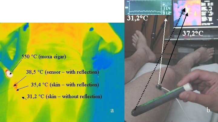
a) Moxa-Zigarre und Infrarot-Wärmestimulation (A burning moxibustion-cigar made of mugwort (length 20 cm, diameter 1.5 cm, Hunan, China) was used for stimulation. This radiates an average temperature of 550°C (measured with FLIR ThermaCAM S65) with an energy maximum ranging from 3 to 3.5 μm). The second stimulation method (infrared heat radiation) was performed with an infrared lamp (EWT, 150 W, Infrared R95E, Philips, Eindhoven, Netherlands). b) Nadel- und Lasernadel-Stimulation (In addition to the first two stimulation methods without actual skin contact, a further standardized acupuncture scheme was used, from which we know it induces proven, reproducible effects in the brain. Following acupoints were stimulated with manual needle and laserneedle acupuncture: Neiguan (Pe.6), Qihai (Ren 6), Zusanli (St.36) and Sanyinjiao (Sp.6). Additional moxibustion was performed at acupoint Qihai. The laserneedles used emitted continuous laser light at a wavelength of 685 nm and output of 30 – 40 mW per laserneedle. Stimulation time was 10 minutes resulting in an energy density of 2.3 kJ/cm2 at each laserneedle and an average total value of 9.2 kJ/cm2 for the entire duration of laser stimulation. Identical acupoints as by needle acupuncture were used. No moxibustion was done during this type of stimulation).
Duchführung der Untersuchung
Eine standardisiertes Vorgehen wurde bei allen Versuchspersonen angewendet: “The volunteers were laid relaxed on a bed for 10 minutes before the examination was started. During this time, the required measurement devices were applied. The first stimulation method was performed with a moxa-cigar. Different areas of the body (left leg, right leg, upper body) were stimulated at a distance of about 10 cm for about 5 minutes. After a resting period, the procedure was repeated using the infrared lamp. In the third part of this study design a standardized acupuncture scheme was used and stimulation with laserneedles, manual needle acupuncture and additional moxibustion of the acupoint Qihai for a duration of 10 minutes was performed. The order of stimulation was selected at random”.
Ergebnisse
“We could not visualize structures which could be connected with possible meridians, as described in Traditional Chinese Medicine, in any of our volunteers. We were able to clearly objectify and quantify technical reflection artifacts; however, indications for a biological correlation could not be found. In the cases when only needle or laserneedle (685 nm) stimulation was used no thermal reflection phenomena were present and also no meridians could be detected.
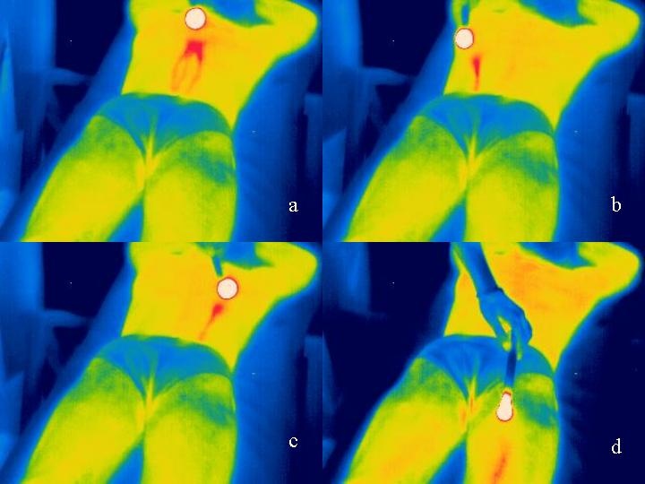
… shows the reflection (mirror-like) artifacts which result from reflection and scattering, in a 30-year-old volunteer. According to the position of the moxa-cigar (circular white area) in dependency with the camera position, reflections showing an optical linear path on the thermogram, can be applied to optional areas of the body.
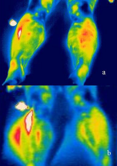
In this manner, typical artifacts from each volunteer can be determined in the “thermographic pictures”. Abbildung 2 shows a further example for this phenomenon. Here, simultaneous measurements in two different wavelength ranges were made using both infrared cameras. While the first camera (7.5 – 13 μm) yielded less reflection artifacts with an all over better optical resolution, the second camera (2 – 5 μm) revealed more distinct artifacts. The energy maximum of the moxa-cigar lies in the range of 3 – 3.5 μm. Since the radiation curve markedly flattens at 7.5 – 13 μm obviously greater reflection occurs within the range of 2 – 5 μm”.
Zusammenfassung
“In conclusion, we note”, so Litscher, “that thermographic methods such as infrared cameras at wavelength ranges of 2 – 5 μm and 7.5 – 13 μm and other High-Tech methods are effective complementary methods in acupuncture research which support demystification of this treatment method. However, the validity of the method for proving meridian structures according to the view of Traditional Chinese Medicine, must be considered critically and analysed scientifically.
In a publication which attempted to visualize meridians and their path using infrared thermography, the authorsdescribed that the left stomach meridian could be visualized during stimulation with a moxa-cigar near the left foot. At the same time, the right spleen meridian should be visualized on the right foot. Based on the foundations of Traditional Chinese Medicine, it is improbable that exactly these two meridians can be activated simultaneously. According to current technical standings, the visualization of energetic paths in the sense of meridians as described in recent literature is not possible using thermography. On the contrary, the supposed thermographic reproduction of meridians is dependent upon physical-technical artifacts caused by thermic reflections.
According to current technical standings and to the method proposed by other authors, the visualization of energetic paths in the sense of meridians seems to be not possible using thermography. Further scientific studies are required regarding the possible visualization of meridians”.
Quellen
Zitate und Bilder aus “Infrared thermography fails to visualize stimulation-induced meridian-like structures”, BioMedical Engineering OnLine 2005, 4:38 doi:10.1186/1475-925X-4-38, http://www.biomedical-engineering-online.com/content/4/1/38